COVID-19 may reduce gray matter volume in brain, small study suggests
- A study found that clients who required oxygen remedy for COVID-19 had lower gray matter quantity in the frontal lobe of their human brain weighed against patients who didn't require supplemental oxygen.
- Reduced gray matter in the frontal lobe also had links with more extreme disability up to 6 months after recovery coming from COVID-19.
- Patients who exactly experienced fever had more affordable gray matter volume found in the temporal lobe weighed against those who didn't.
- However, the analysis was small, thus scientists have to conduct more exploration to confirm the results.
Around 15% of patients hospitalized with COVID-19 experience neurological complications. The symptoms, which happen to be more prevalent in severely ill clients, contain impaired consciousness, confusion, and agitation.
However, the physical ramifications of COVID-19 about the mind are poorly understood.
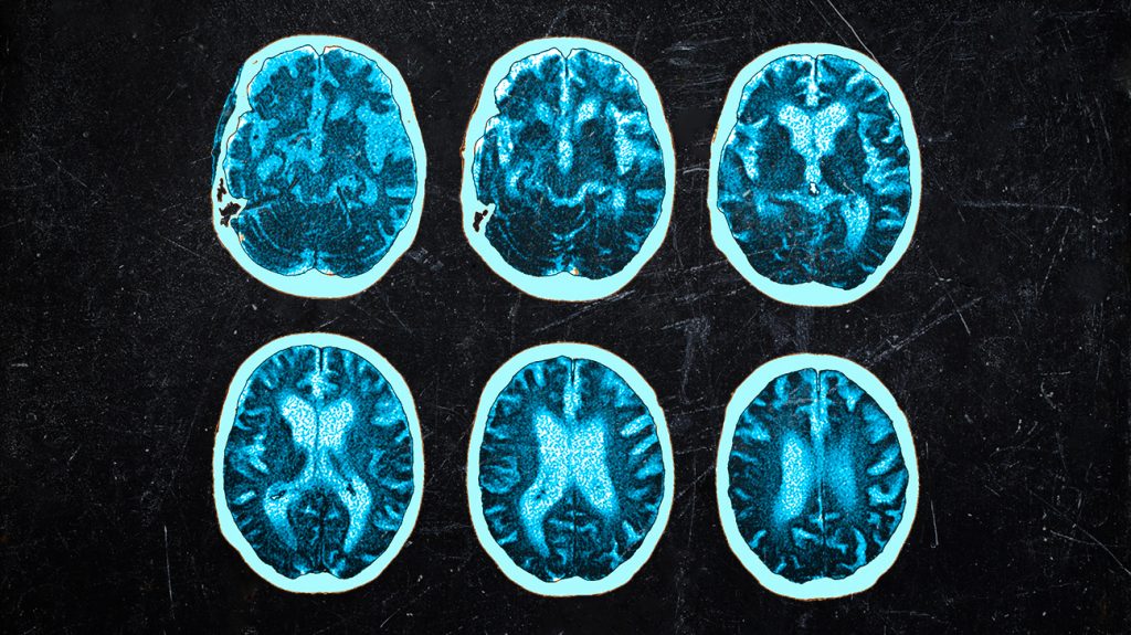
In a fresh study, researchers led by Georgia State University in Atlanta analyzed the CT scans of clients undergoing evaluations for neurological symptoms at a expert hospital in Brescia, Italy.
The team assessed the number of gray matter, which generally comprises the cell bodies of neurons, in the outer layer or cortex of the patients’ brains.
Out of a total of 120 clients, 58 had COVID-19, while 62 didn't. The team matched both teams by age, gender, and other diseases.
While the researchers found no significant dissimilarities in gray subject volume between your two groups, they did get distinctions among the patients with COVID-19.
Those that needed oxygen therapy had reduced gray subject in the frontal lobes of their human brain weighed against those who didn't.
Lower gray matter quantity in the frontal regions likewise had links with more severe disability - that your researchers measured on the modified Rankin scale - up to six months after discharge from a healthcare facility.
Furthermore, patients who knowledgeable a fever during their illness had decreased gray matter in the temporal lobes of their brain weighed against those who didn't have this symptom.
Alterations in mood
The study discovered that reductions in gray matter in the frontal regions of the brain had associations with agitation, suggesting they could underlie the mood changes that recovered patients often experience.
“Neurological difficulties are increasingly documented for patients with COVID-19,” says senior author Vince Calhoun, director of the Tri-institutional Middle for Translational Research in Neuroimaging and Data Science (TReNDS) and professor of psychology at Georgia Talk about.
“A reduction of gray matter in addition has been proven to be present in additional mood disorders, such as schizophrenia, and is probable related to just how that gray subject influences neuron function,” this individual adds.
The study authors suggest that later on, researchers may potentially use alterations in gray subject in the frontal and temporal lobes to look for the long-term prognosis of patients or evaluate treatment plans.
All the associations remained statistically significant even after the experts accounted for preexisting cerebrovascular disease - which impacts blood vessels in the top, diabetes, and hypertension.
They write that the infection may indirectly destruction the discovered brain regions due to fever or insufficient oxygen.
Medical News Today asked the lead author, Kuaikuai Duan, whether a preexisting factor, such as for example having unwanted weight, may have reduced the option of oxygen to affected individuals’ brains and had already caused the observed alterations in gray matter.
“Unfortunately, we did not routinely gather height and weight (to acquire BMI [human body mass index]) for this study,” stated Duan, a graduate exploration assistant at TReNDS and Ph.D. scholar in Georgia Tech’s College of Electrical and Pc Engineering.
“So, we cannot eliminate the probability that the observed differences in gray subject were due to preexisting overweight or obesity.”
Key limitations of the research
The study involved an individual “snapshot” of every patient’s brain, rather than a series of scans spread over time, so it cannot prove that COVID-19 actually caused any of the changes. It simply showed associations between the amount of gray subject and factors such as oxygen supplementation and fever.
In addition, there have been small figures in each subgroup of people, which limits the stability of the findings.
In their paper, the scientists acknowledge various other important limitations.
They write that while hospitals routinely use CT images to diagnose patients, the scans might not exactly adequately capture subtle changes in gray matter volume.
This may clarify why the analysis found no differences in virtually any of the brain regions between patients with COVID-19 and the ones without the disease.
Another explanation may be that some of the changes hadn't occurred when healthcare pros performed the CT scans but were completed 4-5 days, typically, after diagnosis.
The authors remember that two previous studies that did find differences in gray matter between patients with COVID-19 and controls involved MRI scans at around 54 and 90 days after diagnosis.
Finally, the researchers caution that their results may only apply to clients hospitalized with COVID-19 who experience neurological complications.
In the future, they hope to repeat their study on a more substantial group of patients, including diverse sets of COVID-19 patients. They would also like to incorporate info from several types of brain scans.
Source: www.medicalnewstoday.com